Atypical Hyperplasia

Atypical Hyperplasia - What is it?

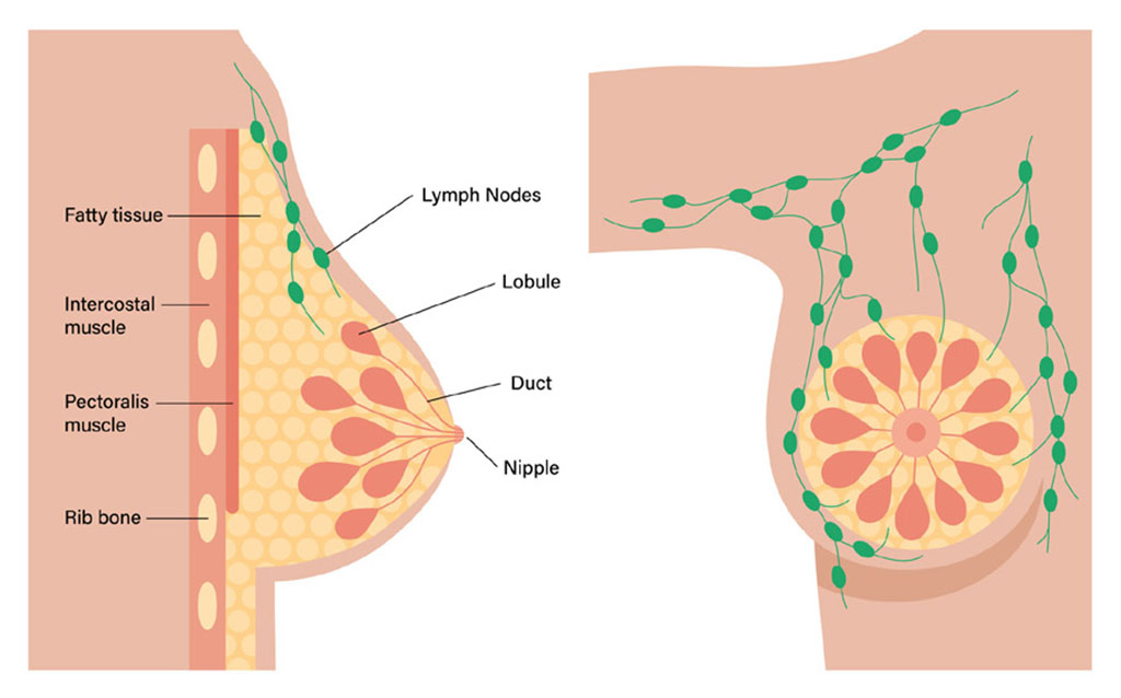
The normal breast is made of ducts (tiny tubes) that end in a group of sacs called lobules. Hyperplasia is a term used when there is growth of cells within the ducts and/or lobules of the breast that is not cancerous. Normally, the ducts and lobules are lined by two layers of cells.

Illustration showing areas of the breast including lobule, duct, nipple and lymph nodes.

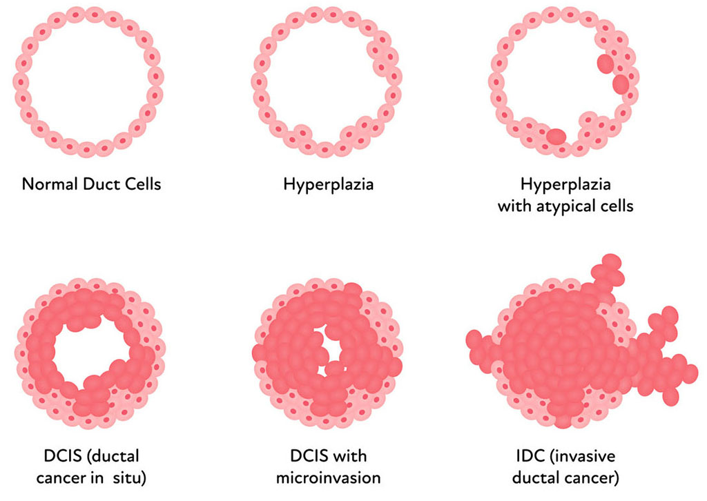
Hyperplasia means that there are more cells than usual and they are no longer lined up in just the two layers. If the growth looks much like the normal pattern under the microscope, the hyperplasia may be called usual. Some growths look more abnormal, and may be called atypical hyperplasia.

Illustration showing breast duct cell types ranging from normal, hyperplasia, and invasive ductal cancer.

The two major patterns of hyperplasia in the breast are ductal hyperplasia (ADH) and lobular hyperplasia (ALH). What makes the hyperplasia ductal or lobular is based more on what the cells look like under the microscope rather than whether the hyperplasia is occurring within the ducts or lobules.

In ADH or ALH, the pattern of growth of cells is abnormal and has some (but not all) of the features of carcinoma in situ, which is a pre-cancer. This means that ADH is not yet a pre-cancer, although it is linked to an increased risk of getting breast cancer later on.

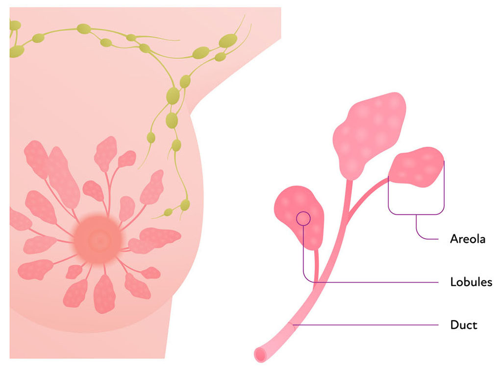
Illustration showing the areola, lobules and duct in a breast.

Treatment Options

A diagnosis of atypical hyperplasia may lead to a surgical biopsy (wide local excision or lumpectomy) to remove all of the affected tissue. The pathologist looks at the larger specimen for evidence of in situ or invasive cancer. Fifteen to twenty percent of needle biopsies that show atypical hyperplasia may have already progressed to carcinoma in situ or invasive cancer at the time of surgery.

If ADH or ALH is found on excisional biopsy after surgery, no additional surgical treatment is needed, but your doctor may recommend taking medicine to help reduce your risk of breast cancer.

Before Your Procedure

Before your procedure, a localization device may be placed to help the surgeon find the area that was biopsied while you are in surgery. This procedure takes place in the Breast Health Center a few days before your surgery and is comparable to what you have already experienced with the biopsy.

Next Steps

Because the condition can progress to cancer, it needs investigation, evaluation, and follow-up.

Whatever your situation, it makes sense to be followed by a doctor with expertise in breast health. Generally, a follow-up physical exam is done at six months and a mammogram may be recommended. After that, most women can resume yearly mammograms; in selected cases doctors may add breast MRI.

For some women, doctors may suggest a hormonal therapy such as tamoxifen, raloxifene, or an aromatase inhibitor to reduce their risk of breast cancer.

Studies have found that most ADH cells test positive for estrogen and progesterone receptors. These treatments have side effects, though, so your individual situation will determine whether their benefits outweigh their risks.

Contact Us

High-Risk Breast Health Clinic

812-376-5710

Medical Office Building South
2325 18th Street, Suite 220
Columbus IN 47201

Hours
Monday- Friday 8am - 5pm

Get Directions Schedule A Mammogram Contact Us Online
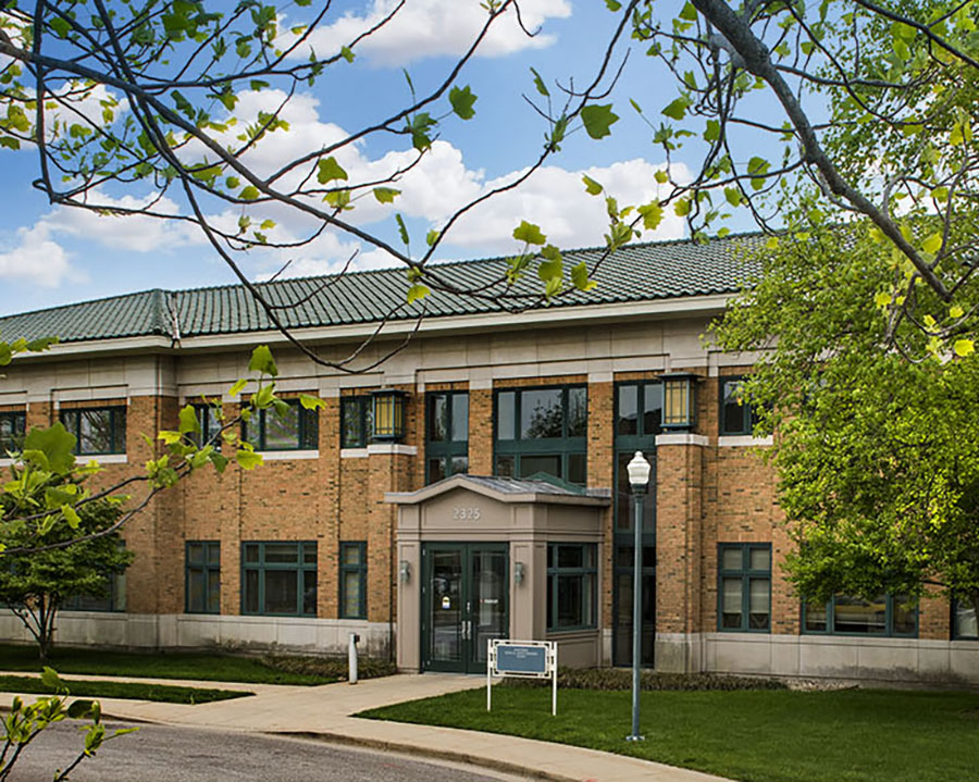
High Risk Breast Health Clinic exterior