
Aortic stenosis is a progressive disease that occurs with a narrowing of the patient’s aortic valve opening.
It primarily happens over time as we age but can also be caused by a birth defect, previous chest radiation, or rheumatic fever. The prevalence of aortic stenosis increases with age. It is estimated that approximately 2.5 million people, or 12.4 percent of the population, in the United States over the age of 75 suffer from aortic stenosis.
Overview of the Disease
Human heart valves are remarkable structures. Normal heart valves have two or three flaps of tissue called leaflets. These tissue-paper thin membranes attached to the heart wall constantly open and close to regulate blood flow (making the sound of a heartbeat).
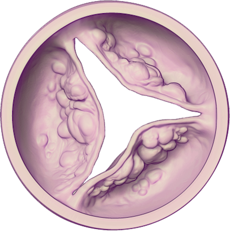 In elderly patients, aortic stenosis is sometimes caused by the buildup of calcium (mineral deposits) on the aortic valve’s leaflets. Over time, the leaflets become stiff, reducing their ability to fully open and close. When the leaflets don’t fully open, a person’s heart must work harder to push blood through the aortic valve to the rest of the body. Eventually, the heart gets weaker, increasing the risk of heart failure (when the heart cannot supply enough blood to the body).
Severe Aortic Stenosis
Aortic stenosis is a progressive disease which means it gets worse over time. It’s typically measured as mild, moderate, or severe aortic stenosis. As a result of the reduced blood flow, the body does not get the oxygen it needs, which may cause symptoms. If a patient has been diagnosed with severe aortic stenosis and is experiencing symptoms, it can be life-threatening and may progress rapidly. However, it’s important to know that Severe Aortic Stenosis may occur with no obvious symptoms. Many patients initially appear asymptomatic, but on closer examination, up to 32 percent exhibit symptoms.
The symptoms listed below are typically associated with severe aortic stenosis but are commonly misunderstood by patients as “normal” signs of aging.
Signs of Severe Aortic Stenosis
You may notice symptoms like:
- Chest pain
- Rapid, fluttering heartbeat
- Trouble breathing or feeling short of breath
- Feeling dizzy or light-headed, even fainting
- Difficulty walking short distances
- Swollen ankles or feet
- Not doing activities you used to enjoy
- Difficulty sleeping or the need to sleep sitting up
Major Risk Factors
Factors associated with aortic valve disease include the following:
- Increasing age
- High blood pressure
- High cholesterol
- Smoking
Severe aortic stenosis is life-threatening, and treatment for this condition is critical. Patients who have developed symptoms from severe aortic stenosis have about a 50 percent chance of living at two years without aortic valve replacement.
Diagnosis
In addition to a physical exam, severe aortic stenosis is diagnosed in several ways, the most common being an echocardiogram, electrocardiogram (EKG), chest X-ray of the patient’s heart, and cardiac catheterization (angiography).
Aortic Valve Replacement Options
Transcatheter Aortic Valve Replacement (TAVR)
TAVR (sometimes called transcatheter aortic valve implantation, or TAVI) is a less invasive procedure than open-heart surgery which allows a new valve to be inserted within the native, diseased aortic valve. The TAVR procedure can be performed using one of many approaches, the most common being the transfemoral approach (through a small incision in the leg). Only a Heart Team can decide which approach is best, based on the patient’s medical condition and other factors.
In preparation for the patient’s transfemoral procedure (or through the upper leg), the patient may be placed under anesthesia. The doctor will make an incision in the leg and insert a short, hollow tube called a sheath. This will allow the doctor to put various devices through the sheath to access the patient’s heart. The new heart valve is placed on the delivery system and compressed onto a balloon to make it small enough to fit through the sheath. Once the delivery system reaches the patient’s diseased valve, the balloon will be inflated with fluid, expanding the new valve into place. The new valve pushes the leaflets of the patient’s diseased valve aside, and the frame of the new valve uses the diseased valve’s leaflets to secure itself into place. The balloon is then deflated and removed. The patient’s doctor will ensure the new valve is working properly before closing up the incision.
Our skilled providers, Dr. Nandu Gourineni, Dr. Ramesh Chillal-Kashinath, Dr. David Hart, and Dr. Gregory Dedinsky, and the Heart & Vascular Team at Columbus Regional Health, are pleased to offer this minimally invasive option to patients so they can receive the best cardiac care close to home.
Contact Southern Indiana Heart and Vascular at 812.379.2020 or visit www.crh.org/tavr for more information.